Your heart isn’t just a muscle; it’s a powerful pump that works tirelessly to circulate blood throughout your entire body. But how does it manage this incredible feat? The answer lies in four remarkable valves that act as one-way doors within your heart. These valves open and close with each beat, ensuring that blood flows in the right direction and at the right time.
Though they may be hidden from view, the heart’s valves are absolutely essential for life. In this article, we’ll examine heart valves in more detail, exploring their names, locations, and how they work together to maintain a healthy and efficient circulatory system.
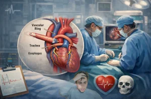
Understanding the Anatomy of the Heart
Before we discuss the specifics of heart valves, let’s familiarize ourselves with the basic structure of this amazing organ.
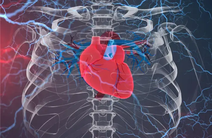
Your heart is a four-chambered muscular organ located slightly to the left of your breastbone. It’s roughly the size of your fist and functions as a tireless pump, circulating blood throughout your body. Think of it as a two-sided pump, with each side playing a distinct role:
- The Right Side: Receives oxygen-poor blood from the body and pumps it to the lungs, where it picks up fresh oxygen.
- The Left Side: Receives oxygen-rich blood from the lungs and pumps it out to the rest of the body, delivering vital oxygen and nutrients to your tissues and organs.
Each side of the heart is further divided into two chambers:
- Atria: The upper chambers that receive incoming blood.
- Ventricles: The lower chambers that pump blood out of the heart.
The separation of oxygen-rich and oxygen-poor blood is essential for efficient circulation. Now, let’s explore the four heart valves that make this intricate system possible.
Prevent heart problems before they start – Schedule a preventive checkup
Contact UsHow Many Valves Does the Human Heart Have?
The human heart contains four valves, each playing a critical role in ensuring the smooth and efficient flow of blood. These valves are strategically positioned at the entrances and exits of the heart’s chambers, acting as one-way gates to prevent blood from flowing backward.
These valves can be classified into two main categories based on their location and structure:
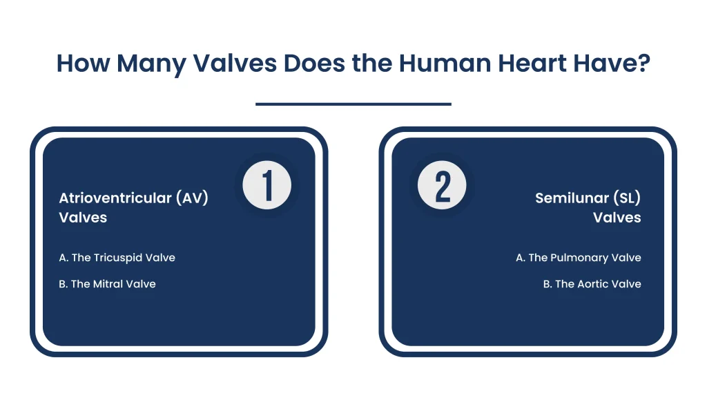
- Atrioventricular (AV) Valves
- Semilunar (SL) Valves
1. Atrioventricular (AV) Valves
These valves are located between the atria (upper chambers) and ventricles (lower chambers) of the heart. They ensure that blood flows from the atria into the ventricles while preventing backflow. The closure of these valves at the start of ventricular contraction (systole) produces the first heart sound (“lub”).
A. The Tricuspid Valve
- Location: Situated between the right atrium (upper chamber) and the right ventricle (lower chamber).
- Function: Acts as a one-way door, allowing blood to flow from the right atrium into the right ventricle while preventing any backflow. When the right atrium contracts, the tricuspid valve opens, and when the right ventricle contracts to pump blood to the lungs, the valve closes to ensure the blood doesn’t flow back into the atrium.
B. The Mitral Valve
- Location: Found between the left atrium (upper chamber) and the left ventricle (lower chamber).
- Function: Similar to the tricuspid valve, the mitral valve acts as a one-way door, permitting blood flow from the left atrium into the left ventricle while preventing backflow. The mitral valve’s proper function is crucial for ensuring that oxygen-rich blood reaches the body’s tissues and organs.
2. Semilunar (SL) Valves
These valves are situated at the base of the major arteries leaving the ventricles. They ensure that blood flows out of the ventricles and into the arteries while preventing backflow into the heart. The closure of these valves at the beginning of ventricular relaxation (diastole) produces the second heart sound (“dub”).
A. The Pulmonary Valve
- Location: Positioned between the right ventricle and the pulmonary artery, the vessel that carries oxygen-poor blood to the lungs.
- Function: Like the tricuspid valve, the pulmonary valve opens to allow blood to flow from the right ventricle into the pulmonary artery and then closes to prevent backflow into the ventricle. This valve plays a crucial role in directing blood to the lungs, where it picks up vital oxygen.
B. The Aortic Valve
- Location: Situated between the left ventricle and the aorta, the largest artery in the body, which carries oxygen-rich blood to the rest of the body.
- Function: The aortic valve opens to allow the left ventricle to pump oxygenated blood into the aorta, and then it closes tightly to prevent any backflow into the ventricle. This valve’s efficient operation is essential for maintaining a steady supply of oxygenated blood throughout the body.
Prevent heart problems before they start – Schedule a preventive checkup
Contact UsThe Structure of Heart Valves: Leaflets and More
Heart valves aren’t just simple openings; they are complex structures designed to facilitate the one-way flow of blood. Each valve consists of several key components working in harmony:
A. Leaflets (Cusps)
Leaflets are thin, flexible flaps of tissue that open and close like gates, allowing blood to flow through the valve in the correct direction while preventing backflow. The specific number and shape of leaflets vary among the valves:
- Tricuspid Valve Leaflets:
- Anterior Leaflet: The largest leaflet, located towards the front of the heart.
- Posterior Leaflet: Positioned towards the back of the heart.
- Septal Leaflet: Attached to the septum, the wall that separates the heart’s right and left sides.
- Pulmonary Valve Leaflets (Cusps):
- Left Cusp: Forms the left side of the valve.
- Right Cusp: Forms the right side of the valve.
- Anterior Cusp: Situated towards the front of the heart.
- Mitral Valve Leaflets:
- Anterior Leaflet: A larger leaflet that constitutes most of the valve’s opening.
- Posterior Leaflet: A smaller leaflet that completes the valve’s structure.
- Aortic Valve Leaflets (Cusps):
- Left Cusp: Forms the left side of the valve.
- Right Cusp: Forms the right side of the valve.
- Non-coronary Cusp: Positioned neither towards the left nor the right coronary artery.
B. Chordae Tendineae and Papillary Muscles: The Valve’s Support System
Leaflets alone cannot ensure proper valve closure. They rely on two additional structures for support:
- Chordae Tendineae: These are thin, strong, tendon-like cords that attach the edges of the leaflets to the papillary muscles. They act like tethers, preventing the leaflets from inverting or prolapsing into the atrium when the ventricles contract.
- Papillary Muscles: These are small muscles that project inward from the walls of the ventricles. When the ventricles contract, the papillary muscles also contract, pulling on the chordae tendineae and helping to close the valve leaflets tightly.
The coordinated action of leaflets, chordae tendineae, and papillary muscles ensures that blood flows smoothly through the heart in the correct direction and that no backflow occurs.
D. The Valve Annulus
The valve annulus is a fibrous ring that encircles the base of each valve. It provides structural support to the leaflets and helps maintain the valve’s shape and integrity.
Final Words
The heart’s four valves work tirelessly to ensure the smooth and efficient circulation of blood throughout your body. Their precise coordination and one-way flow mechanism are essential for maintaining a healthy cardiovascular system. Understanding the structure and function of these valves sheds light on the complex workings of the human heart and the vital role it plays in sustaining life.