Stress tests are one of the most common ways doctors evaluate how well your heart works when it’s pushed because many heart problems show up during stress (exercise or medicine-induced stress) even when you feel okay at rest. These tests help uncover issues like reduced blood flow to the heart and can guide next steps for treatment or prevention.
A nuclear stress test is a special type of stress test that combines stress on the heart with imaging that shows how blood flows through the heart muscle. It can offer more precise details than an ECG-only stress test, especially in certain people.
What Is a Stress Test?
A stress test checks how your heart responds when it has to work harder than usual. Think of it as a “performance test” for your heart, either by walking on a treadmill or by receiving a medication that mimics the effects of exercise.
A stress test typically measures:
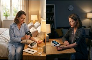
- Heart rhythm and electrical activity (via ECG/EKG)
- Heart rate and blood pressure response
- Symptoms during exertion (chest pain, shortness of breath, dizziness, fatigue)
- In some tests, heart muscle movement (echo) or blood flow (nuclear imaging)
Doctors often order a stress test if you have:
- Chest pain or pressure
- Shortness of breath with activity
- Unexplained fatigue, dizziness, or palpitations
- Risk factors for coronary artery disease (high blood pressure, diabetes, smoking, high cholesterol, family history)
- An abnormal ECG or concerning findings from another test
- A need to assess heart function before certain surgeries
Types of Stress Test
Different stress tests answer slightly different questions. Your doctor chooses the type based on symptoms, risk level, ability to exercise, and what information is needed.
Exercise Stress Test (Treadmill/ECG)
This is the most basic stress test. You walk on a treadmill (or pedal a bike) while the medical team monitors your ECG, heart rate, and blood pressure.
What’s best for:
- Detecting ECG changes that suggest reduced blood flow
- Assessing exercise capacity and symptom triggers
Limitations:
- Some people can’t exercise enough for accurate results
- ECG changes can be more complex to interpret in certain conditions (e.g., baseline ECG abnormalities)
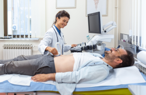
Stress Echocardiogram
A stress echo uses ultrasound imaging of your heart, taken at rest and after exercise (or medication-induced stress). The images show how well heart muscle segments contract under stress.
What’s best for:
- Detecting areas of poor heart muscle movement due to reduced blood supply
- Evaluating valve function alongside stress response
Limitations:
- Image quality can vary depending on body structure and lung interference
- Doesn’t directly “map” blood flow like nuclear imaging
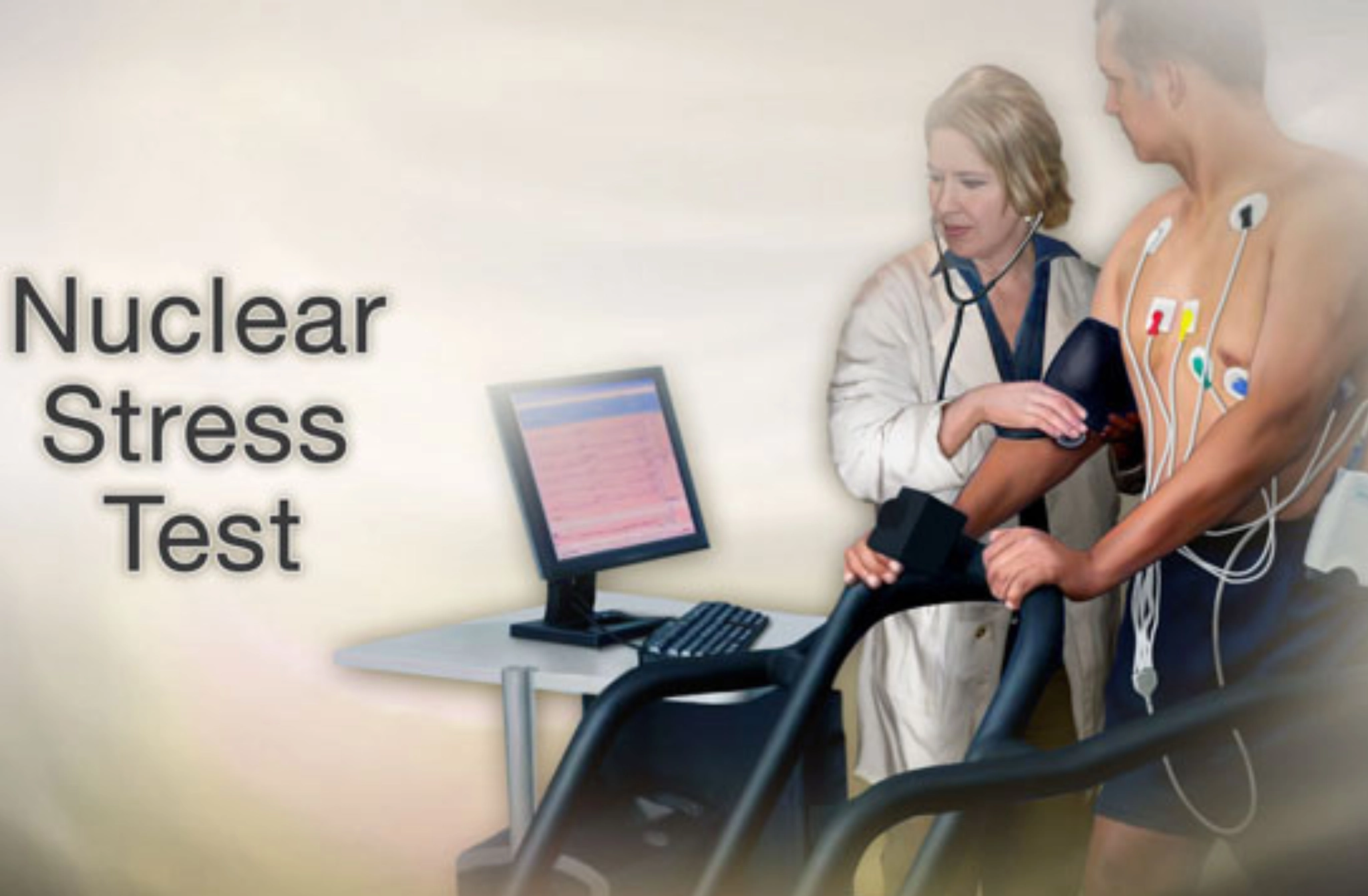
Nuclear Stress Test (Myocardial Perfusion Imaging)
A nuclear stress test uses a small amount of a radioactive tracer and a special camera to see blood flow to the heart muscle at rest and during stress. Areas with reduced blood flow may appear as “defects” on the scan.
What’s best for:
- Identifying reduced blood flow (ischemia)
- Distinguishing older damage (scar) from active blood flow problems
- Risk stratification in many patients with moderate-to-high risk
Pharmacologic Stress Test
If you can’t exercise enough (due to joint pain, mobility limitations, lung disease, or other reasons), a medication can simulate the heart’s response to exertion by increasing blood flow demands or dilating coronary vessels.
Common approach:
- Medication-induced stress + imaging (often nuclear, sometimes echo)
Good for:
- People are unable to reach the target heart rate with exercise
Purpose of Stress Test
Stress tests help doctors answer: Does your heart get enough blood and oxygen when it’s working hard?
Common goals include:
- Detect reduced blood flow (ischemia):
When coronary arteries are narrowed, the heart muscle may not receive enough blood during exertion.
- Evaluate chest pain or shortness of breath:
Stress testing helps determine whether symptoms may relate to the heart.
- Check the effectiveness of treatment:
After stents, bypass surgery, or medication changes, stress tests can show whether blood flow and risk have improved.
- Determine safe exercise level or surgical clearance:
Some patients need cardiac evaluation before major surgery or before starting a new exercise routine.
Prevent heart problems before they start – Schedule a preventive checkup
Contact UsWho Should and Should Not Have This Test
Who Should Consider It (Common Candidates)
A nuclear stress test may be considered when there is concern about coronary artery disease or when a more detailed picture of blood flow is needed.
Common candidates include:
- People with chest pain, pressure, tightness, or discomfort (especially with exertion)
- People with shortness of breath or exercise intolerance
- Those with known coronary artery disease or prior heart attack
- People with higher risk factors, such as:
- Diabetes
- High blood pressure
- High cholesterol
- Smoking history
- Strong family history of early heart disease
- Patients with an abnormal ECG or results from prior tests that need clarification
Who May Not Be Eligible (or Needs Caution)
Not everyone is a good fit for a nuclear stress test, and sometimes the test is delayed or replaced with another.
Situations requiring caution include:
- Pregnancy (radiation concerns)
- Specific heart rhythm issues that make interpretation difficult or increase risk
- Severe uncontrolled hypertension
- Acute illness (fever, infection) or unstable symptoms (ongoing chest pain at rest, severe shortness of breath)
- Recent significant cardiac events where immediate evaluation may be needed differently
How to Prepare for It
Preparation can vary by clinic and by the medication used for stress, so follow the instructions you’re given. Below are standard guidelines.
24 Hours Before
You may be asked to:
- Avoid caffeine (coffee, tea, energy drinks, soda, chocolate, certain headache medicines) because caffeine can interfere with some stress medications.
- Avoid smoking or nicotine products if instructed, since they can affect heart rate and blood vessels.
Day of the Test
Typical recommendations:
- Wear comfortable clothes and walking shoes (even if you’ll likely receive medication, some centers still use a treadmill if possible).
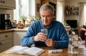
- Bring a current medication list (names, doses, and timing).
- Follow fasting instructions if provided (some centers ask you not to eat for a few hours beforehand).
- If you have diabetes, ask specifically how to manage insulin or oral medications that morning.
Medications to Discuss
Do not stop medications unless your clinician instructs you to. However, some medications may be adjusted before a stress test, such as:
- Beta blockers (can limit heart rate response)
- Nitrates (can affect symptoms and blood flow response)
- Diabetes medications (due to fasting)
- Inhalers (especially if you have asthma/COPD and might receive certain stress medications)
Always clarify medication instructions directly with your care team, because guidance depends on your specific health situation and the type of stress protocol used.
What Happens Before and After a Stress Test
Before the Test
Most stress testing visits start with:
- Check-in and paperwork
- A review of symptoms and medical history
- Vitals (blood pressure, heart rate, oxygen level)
- Placement of ECG leads on your chest
- Starting an IV line (especially for nuclear stress tests, since tracer is injected)
- For nuclear stress testing, baseline (resting) imaging may be performed before the stress portion or after tracer injection at rest.
After the Test
After the stress portion and imaging:
- You’ll usually have a short monitoring period to ensure heart rate and blood pressure stabilize.
- You may be advised to drink water to help flush the tracer from your body.
- Many people can return to normal activities the same day, depending on how they feel and what medication was used.
When to call a doctor after the test:
Seek medical help if you experience severe or worsening chest pain, fainting, severe shortness of breath, or symptoms that feel unusual for you.
What to Expect During a Nuclear Stress Test
A nuclear stress test is often done in phases so the imaging can compare blood flow at rest versus under stress.
Step 1: Resting Scan
- You’ll receive a tracer injection through your IV.
- Then there is typically a waiting period (often 15–60 minutes) so the tracer circulates and is taken up by the heart muscle.
- You lie on a table while a special camera captures images of your heart. You’ll need to stay pretty still, but the procedure is usually painless.
Step 2: Stress Phase
Stress is created in one of two ways:
- Treadmill exercise:
You walk as the speed and incline increase gradually. The goal is to reach a target heart rate.
- Medication-induced stress:
If you can’t exercise enough, you’ll receive medicine that increases heart workload or changes blood flow patterns to mimic exercise effects.
During stress, the team monitors:
- Heart rhythm (ECG)
- Blood pressure
- Symptoms (chest discomfort, shortness of breath, dizziness, fatigue)
At peak stress, a second dose of tracer may be injected (depending on protocol).
Step 3: Stress Scan
- After stress and tracer injection, you may wait again briefly.
- The camera takes a second set of images to evaluate stress perfusion (blood flow under stress).
How long does it take?
Total time varies by clinic and protocol. Many nuclear stress tests take 2 to 4 hours in total, sometimes longer if multiple waiting periods are used or if the clinic uses a two-part protocol.
Does a Nuclear Stress Test Use Radiation?
Yes.
A nuclear stress test uses a radioactive tracer so the camera can visualize how blood flows to the heart muscle. The tracer emits radiation that the imaging system detects to create detailed pictures.
The dose is carefully controlled, and these tests are used because the diagnostic value can be significant, particularly when determining whether reduced blood flow is present and how serious it may be.
How Much Radiation Is in a Nuclear Stress Test?
Radiation exposure varies depending on the tracer and protocol used. Some centers use lower-dose methods or modern equipment that reduces exposure while maintaining image quality.
Nuclear Stress Test Radiation: What Affects the Dose?
Several factors can change your total radiation dose, including:
- Type of tracer used (different tracers have different dose profiles)
- One-day vs two-day protocol (dose can be split depending on scheduling and patient factors)
- Equipment and clinic protocols (newer cameras and optimized techniques can reduce dose)
- Patient body size (image quality needs can influence dose)
Is the Radiation From a Nuclear Test Harmful?
Yes.
Radiation exposure carries a theoretical long-term risk, especially with repeated exposures over time. That said, in medical imaging, radiation is used when clinicians believe the benefits outweigh the risks, for example, when diagnosing or ruling out coronary artery disease, it can prevent a heart attack or guide life-saving treatment.
Special considerations include:
- Pregnancy: Nuclear testing is usually avoided unless absolutely necessary.
- Repeated imaging: If you’ve had many CT scans or nuclear tests, ask whether alternative tests (like stress echo or MRI) could be appropriate.
For most people, a single nuclear stress test’s radiation exposure is considered low enough that the diagnostic information can be worth it, especially when symptoms or risk level suggest meaningful heart disease may be present.
Risks and Benefits of This Test
Benefits
A nuclear stress test can provide high-value information, such as:
- Detecting blocked or narrowed arteries by revealing reduced blood flow
- Helping guide treatment decisions (medication optimization, angiography, stenting, lifestyle interventions)
- Risk stratification helps determine whether someone is low, intermediate, or high risk for future cardiac events
- Blood flow problems that might be reversible (treatable ischemia)
- Areas of older damage/scar
Risks (Rare but Possible)
While nuclear stress tests are generally safe, potential risks include:
- Allergic reaction to the tracer (rare)
- Arrhythmia during stress (usually short-lived)
- Symptoms during stress: chest pain, dizziness, nausea, shortness of breath, and blood pressure changes
- Radiation exposure (small risk, weighed against clinical benefit)
The test is supervised by trained staff with safety protocols in place.
Prevent heart problems before they start – Schedule a preventive checkup
Contact UsResults and Follow-Up
Results usually compare rest images with stress images.
Common result patterns include:
Normal perfusion:
Blood flow looks adequate at rest and under stress—often reassuring.
Abnormal perfusion:
One or more areas show reduced tracer uptake, suggesting reduced blood flow or prior damage.
Two terms you might hear:
Reversible defect:
Blood flow looks reduced during stress but improves at rest. This can suggest ischemia, a potentially treatable blood flow limitation.
Fixed defect:
Reduced tracer uptake both at rest and stress. This can suggest scar tissue from a prior heart attack (though interpretation depends on context).
Some reports also include ejection fraction (EF), a measure of how effectively the heart pumps blood. EF is one piece of the overall picture, not the only decision-maker.
What Happens Next?
Next steps depend on symptoms, risk factors, and severity. Your clinician may recommend:
- Lifestyle changes (exercise plan, nutrition, smoking cessation, weight management)
- Medications (cholesterol-lowering therapy, blood pressure meds, anti-anginals, diabetes optimization)
- Additional testing, such as:
- CT coronary angiography (a CT-based look at coronary arteries)
- Cardiac catheterization (coronary angiography) fora more direct evaluation
- A cardiology follow-up (often within days to weeks, sooner if results are concerning)
If your test was ordered for pre-surgery clearance or to evaluate symptoms, your doctor will explain how results affect timing and next steps.
Conclusion
A nuclear stress test is a powerful tool for evaluating how well your heart receives blood flow during stress compared with rest. It can help identify reduced blood supply, clarify symptoms like chest pain or shortness of breath, and guide treatment decisions. While it does involve radiation, the dose is controlled, and the test is typically recommended when the expected diagnostic benefits outweigh the risks.
If you’re scheduled for a nuclear stress test, ask your provider about preparation steps, medication instructions, estimated radiation dose, and whether alternative tests might be appropriate for your situation.
FAQs
Q1.How long does a nuclear stress test take?
Ans: Most nuclear stress tests take about 2 to 4 hours, sometimes longer, depending on waiting periods and the clinic’s protocol.
Q2.Can I drive home after a nuclear stress test?
Ans: Many people can drive themselves home. However, if you receive certain medications or feel lightheaded afterward, you may prefer someone accompany you. Follow your clinic’s instructions.
Q3.What should I eat after a nuclear stress test?
Ans: Unless your clinic says otherwise, you can usually eat normally afterward. Many centers encourage hydration (water) to help your body clear the tracer.
Q4.Can I take my medications before the test?
Ans: Some medications may need to be paused, but only if your clinician instructs you. Bring your medication list and confirm instructions ahead of time.
Q5.Is a nuclear stress test painful?
Ans: The test is usually not painful. You may feel brief discomfort from the IV insertion, treadmill exertion, or temporary side effects from stress medication (like flushing or shortness of breath).
Q6.How soon will I get my results?
Ans: Some clinics provide preliminary impressions the same day, but formal results are often available within 1–3 business days. Timing depends on how quickly the images are interpreted and reported.